Diagnostic Imaging
When your pet is unwell, they may need further investigation and biopsy samples taken to obtain a diagnosis. We have state of the art equipment which allows our highly trained vets to examine inside the body, visualise organs and take samples without invasive surgery.
Radiography
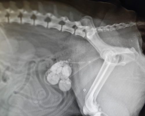
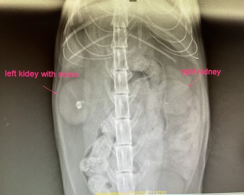
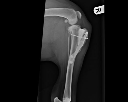
Our digital x-ray machine and moveable table lets us take the perfect image in just a few seconds. This means we don’t have to move the patient between images and the time under anaesthetic is kept to a minimum. It is most useful for looking at bones, foreign objects and abnormalities in the chest and abdomen.
Ultrasound
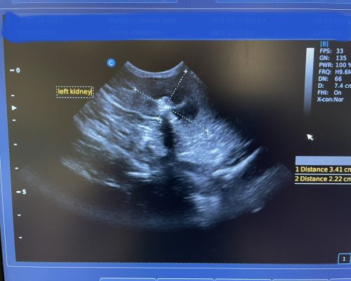
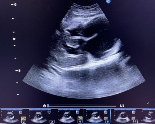
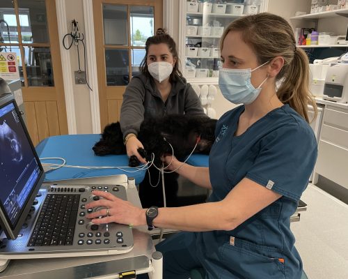
This non-invasive and non-painful method of examining organs uses ultrasonic sound waves from a small probe placed against the skin to create images of internal organs. We can measure their size and shape as well as detecting abnormalities within the tissues and using it to guide sample collection. It is most useful for abdominal organs including pregnancy diagnosis and also a very important part of investigating heart disease.
Our vets are all experienced at abdominal ultrasound and Louise has undertaken extra training in scanning hearts.
Find Us
Opening Hours
- Monday - Friday8:30 - 18:30
- SaturdayClosed
- SundayClosed
- Bank HolidaysClosed
Opening Hours
- Monday - Friday8:30 - 18:30
- Saturday8:30 - 14:00
- SundayClosed
- Bank HolidaysClosed
Emergencies
If you have any concerns about your pet's health while we are closed, please call our our of hours emergency providers, Medivet 24 Hour Wokingham on:
01189 790 551
Book an
appointment
We know how busy life can be. Online appointment booking available 24/7.
Book appointmentEmergencies
If you have any concerns about your pet's health while we are closed, please call our our of hours emergency providers, MiNightVet on:
0118 973 3466


